Animal Cell Diagram Centrioles - Centrioles Vector Images 48 / Gall and etienne de harven in the 1950s.. What is an example of a centriole? However mostly animal cells have them. Nov 13, 2015 · centrioles. Although, several recent types of research have explained that the cell which does not have a centriole (surgically removed through laser) can function without it in the g1 level of. What are facts about the centriole?
The centriole is an organelle inside cells. They lack a limiting membrane and dna or rna and occur in most algal cells (a notable exception being red algae), moss cells, some fern cells, and most animal cells. Found only in animal cells, these paired organelles are typically located together near the nucleus in the centrosome, a granular mass that serves as an organizing center for microtubules. Centrioles are constructed of microtubules. For this reason, they are located near the nucleus.
Diagram A Animal Cell Diagram Full Version Hd Quality Cell Diagram Ritualdiagrams Itfpontederadevitalia It from 4.bp.blogspot.com Each centriole has nine bundles of microtubules which are hollow tubes that give organelles their shape. Aug 22, 2020 · centrioles definition structure functions and diagram centrioles play a notable role in cell division. The structure of duplication of centrioles was first given by joseph g. Found only in animal cells, these paired organelles are typically located together near the nucleus in the centrosome, a granular mass that serves as an organizing center for microtubules. They lack a limiting membrane and dna or rna and occur in most algal cells (a notable exception being red algae), moss cells, some fern cells, and most animal cells. The centriole is an organelle inside cells. The pair of centrioles is often called diplosome. More images for animal cell diagram centrioles »
More images for animal cell diagram centrioles »
More images for animal cell diagram centrioles » Apart from cell division, centrioles are also involved in the formation of cilia and flagella and thus contribute to cell movement. Each centriole has nine bundles of microtubules which are hollow tubes that give organelles their shape. The centriole is an organelle inside cells. What is an example of a centriole? However mostly animal cells have them. Gall and etienne de harven in the 1950s. The pair of centrioles is often called diplosome. What are facts about the centriole? Found only in animal cells, these paired organelles are typically located together near the nucleus in the centrosome, a granular mass that serves as an organizing center for microtubules. Although, several recent types of research have explained that the cell which does not have a centriole (surgically removed through laser) can function without it in the g1 level of. Centrioles are found as single structures in cilia and flagella in animal cells and some lower plant cells. Centrioles are constructed of microtubules.
Centriole helps in organizing the mitotic spindle and complete the process of cytokinesis. What are facts about the centriole? Within the centrosome, the centrioles are positioned so that they are at right angles to each other, as illustrated in figure 1. The structure of duplication of centrioles was first given by joseph g. Centrioles are constructed of microtubules.
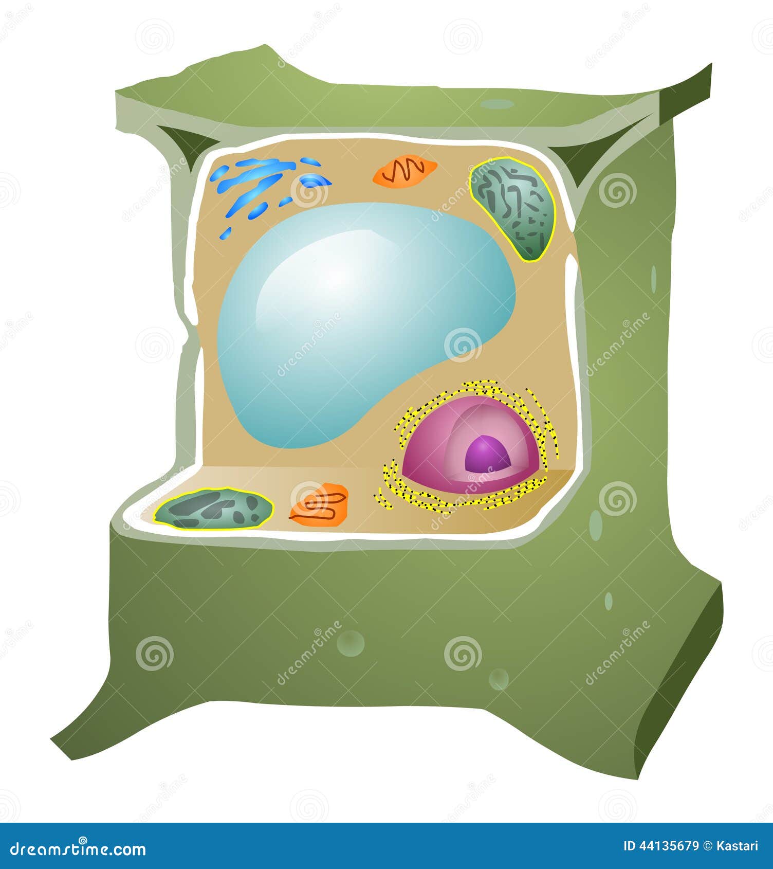
Plant Cell Stock Vector Illustration Of Organelles Biology 44135679 from thumbs.dreamstime.com However mostly animal cells have them. The centriole is an organelle inside cells. For this reason, they are located near the nucleus. However, centrioles were believed to be necessary for the formation of the mitotic spindle in the animal cell. The pair of centrioles is often called diplosome. Found only in animal cells, these paired organelles are typically located together near the nucleus in the centrosome, a granular mass that serves as an organizing center for microtubules. Each centriole has nine bundles of microtubules which are hollow tubes that give organelles their shape. Apart from cell division, centrioles are also involved in the formation of cilia and flagella and thus contribute to cell movement.
However mostly animal cells have them.
How many centrosomes are there in animal cells? In the cell, centrioles aid in cell division by facilitating the separation of chromosomes. Nov 13, 2015 · centrioles. The pair of centrioles is often called diplosome. Centrioles occur as paired cylindrical organelles together with pericentriolar material (pcm) in the centrosome of an animal cell. Each centriole has nine bundles of microtubules which are hollow tubes that give organelles their shape. Although, several recent types of research have explained that the cell which does not have a centriole (surgically removed through laser) can function without it in the g1 level of. For this reason, they are located near the nucleus. Gall and etienne de harven in the 1950s. However mostly animal cells have them. What is an example of a centriole? • centrioles • cytoskeleton • has dna • no cell wall • fixed shape • shape can change • cytoplasm • ribosomes • centrioles • cytoskeleton • has dna • no cell wall • fixed shape • shape can change • makes more cells through mitosis/ meiosis plant cells animal cells venn diagram of plant and animal cells Where are the centrioles located in the animal cell?
How many centrosomes are there in animal cells? Centriole helps in organizing the mitotic spindle and complete the process of cytokinesis. Within the centrosome, the centrioles are positioned so that they are at right angles to each other, as illustrated in figure 1. The pair of centrioles is often called diplosome. Gall and etienne de harven in the 1950s.
Centriole Number Control In Specialized Cell Types A Multiple Download Scientific Diagram from www.researchgate.net The centriole is an organelle inside cells. Where are the centrioles located in the animal cell? Each centriole has nine bundles of microtubules which are hollow tubes that give organelles their shape. Centrioles are found as single structures in cilia and flagella in animal cells and some lower plant cells. However, centrioles were believed to be necessary for the formation of the mitotic spindle in the animal cell. In the cell, centrioles aid in cell division by facilitating the separation of chromosomes. Found only in animal cells, these paired organelles are typically located together near the nucleus in the centrosome, a granular mass that serves as an organizing center for microtubules. Centriole helps in organizing the mitotic spindle and complete the process of cytokinesis.
More images for animal cell diagram centrioles »
Centriole helps in organizing the mitotic spindle and complete the process of cytokinesis. The centriole is an organelle inside cells. Nov 13, 2015 · centrioles. • centrioles • cytoskeleton • has dna • no cell wall • fixed shape • shape can change • cytoplasm • ribosomes • centrioles • cytoskeleton • has dna • no cell wall • fixed shape • shape can change • makes more cells through mitosis/ meiosis plant cells animal cells venn diagram of plant and animal cells However mostly animal cells have them. Aug 22, 2020 · centrioles definition structure functions and diagram centrioles play a notable role in cell division. Although, several recent types of research have explained that the cell which does not have a centriole (surgically removed through laser) can function without it in the g1 level of. Within the centrosome, the centrioles are positioned so that they are at right angles to each other, as illustrated in figure 1. Centrioles are constructed of microtubules. Apart from cell division, centrioles are also involved in the formation of cilia and flagella and thus contribute to cell movement. Where are the centrioles located in the animal cell? What is an example of a centriole? The pair of centrioles is often called diplosome.
Share :
Post a Comment
for "Animal Cell Diagram Centrioles - Centrioles Vector Images 48 / Gall and etienne de harven in the 1950s."


Post a Comment for "Animal Cell Diagram Centrioles - Centrioles Vector Images 48 / Gall and etienne de harven in the 1950s."